Home » Uncategories » Human Bone Anatomy : The Perfect Human Face: Anatomy of Facial Bones - Bone marrow is the soft, flexible connective tissue within bone cavities.
Human Bone Anatomy : The Perfect Human Face: Anatomy of Facial Bones - Bone marrow is the soft, flexible connective tissue within bone cavities.
Human Bone Anatomy : The Perfect Human Face: Anatomy of Facial Bones - Bone marrow is the soft, flexible connective tissue within bone cavities.. Its lower end helps create the knee joint. Disarticulated human skeleton model for anatomy 67 inch high, full size skeleton models with poster, skull, bones, articulated hand & foot, for anatomy art halloween decor 4.8 out of 5 stars 44 $87.85 $ 87. Although the tailbone is considered vestigial (or no longer necessary) in the human body, it does have some function in the pelvis. The skeletal system also provides attachment points for muscles to allow movements at the joints. Chris the skeletal system is made up of some 206 bones in adults which provides form for the soft tissues of the body, protection and a hard lever structure for the muscles.
The frontal bone is a flat bone. Its lower end helps create the knee joint. Many muscles that move the trunk and legs, such as our abdominal muscles, attach to the hip bones. The femur is an example of a long bone. Beyond these similarities, however, lie some profound differences.
Images 04. Skeletal System | Basic Human Anatomy from brooksidepress.org It has every bone and organ in the human body. For anatomy students and medical students, it's important to note that this skeleton's right forearm is rotated forward to show how the arm bones look from a different angle. The skeletal system also provides attachment points for muscles to allow movements at the joints. Posted on may 28, 2014 by admin. Disarticulated human skeleton model for anatomy 67 inch high, full size skeleton models with poster, skull, bones, articulated hand & foot, for anatomy art halloween decor 4.8 out of 5 stars 44 $87.85 $ 87. The skeleton of an adult human is made up of 206 bones of many different shapes and sizes. Although the tailbone is considered vestigial (or no longer necessary) in the human body, it does have some function in the pelvis. The diaphysis and the epiphysis.
Its lower end helps create the knee joint.
This framework consists of many individual bones and cartilages. Bone structure consists of a number of layers. The femur is an example of a long bone. A true and totally 3d free app for learning human anatomy with position quiz, built on an advanced interactive 3d touch interface. For anatomy students and medical students, it's important to note that this skeleton's right forearm is rotated forward to show how the arm bones look from a different angle. Its lower end helps create the knee joint. The skeleton of an adult human is made up of 206 bones of many different shapes and sizes. Bones of the human skeletal system are categorized by their shape and function into five types. This structure encloses and protects the principal organs of circulation and respiration, the heart and the lungs, and is the base to which the upper limbs are attached. In addition, the broad hip bones provide protection to the delicate internal organs of the pelvis, such as the intestines, urinary bladder, and uterus. Typical of mammalian structure, the human body shows such characteristics as hair, mammary glands, and highly developed sense organs. Human skeleton, the internal skeleton that serves as a framework for the body. Posted on may 28, 2014 by admin.
There also are bands of fibrous connective tissue —the ligaments and the tendons —in intimate relationship with the parts of the skeleton. Bone zygomatic bone maxilla mandible nasal bones perpendicular plate of ethmoid nasal conchae note the nasal bones only make up a small portion of the bridge of the nose, most of the external nose is cartilage. Produces a collection of high quality casts of the human skeleton models for educators and practitioners in medicine, physical therapy, physical anthropology, comparative anatomy, and biomechanics. 17 anatomy of the eye macular degeneration. 15 anatomy of the ear swimmer's ear.
Pelvis bones and the ligaments front on and rear view ... from i.pinimg.com 15 anatomy of the ear swimmer's ear. Red marrow and yellow marrow. It is important for bones to be strong to support our body weight. Browse 31,371 human skeleton anatomy stock photos and images available, or search for human bones or human anatomy to find more great stock photos and pictures. Gross anatomy of bone the structure of a long bone allows for the best visualization of all of the parts of a bone (link). Added together, your bones make up about 15% of your body weight. Here we explain the anatomy of bone and the function of each part. In addition, the broad hip bones provide protection to the delicate internal organs of the pelvis, such as the intestines, urinary bladder, and uterus.
Bone zygomatic bone maxilla mandible nasal bones perpendicular plate of ethmoid nasal conchae note the nasal bones only make up a small portion of the bridge of the nose, most of the external nose is cartilage.
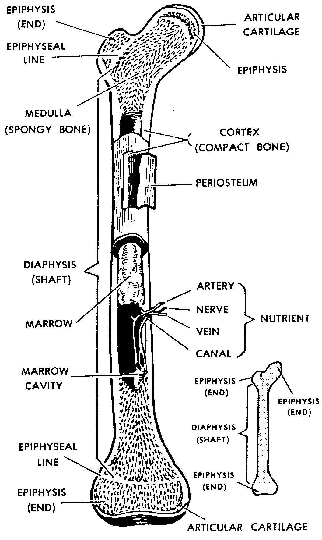
Posted on may 28, 2014 by admin. Human skeleton, the internal skeleton that serves as a framework for the body. The skeleton of the human thorax or chest is like a basket or cage composed of cartilage and bone. Browse 31,371 human skeleton anatomy stock photos and images available, or search for human bones or human anatomy to find more great stock photos and pictures. These include the periosteum, compact bone, spongy bone and an inner core of bone marrow. A component of the lymphatic system, bone marrow functions primarily to produce blood cells and to store fat.bone marrow is highly vascular, meaning that it is richly supplied with a large number of blood vessels.there are two categories of bone marrow tissue: The skeleton of an adult human is made up of 206 bones of many different shapes and sizes. Each bone is a complex living organ that is made up of many cells, protein fibers, and minerals. The human skeletal system consists of all of the bones, cartilage, tendons, and ligaments in the body. It provides structure to the body, and each bone has a distinct purpose. 17 anatomy of the eye macular degeneration. Posted in diagrams | tagged all bones, human skeleton, skelet, skeleton human eye featured. For anatomy students and medical students, it's important to note that this skeleton's right forearm is rotated forward to show how the arm bones look from a different angle.
There also are bands of fibrous connective tissue —the ligaments and the tendons —in intimate relationship with the parts of the skeleton. This is not the standard positioning found in most anatomy diagrams, so keep in mind that in most diagrams, both arms are positioned like this skeleton's left arm. Disarticulated human skeleton model for anatomy 67 inch high, full size skeleton models with poster, skull, bones, articulated hand & foot, for anatomy art halloween decor 4.8 out of 5 stars 44 $87.85 $ 87. In addition, the broad hip bones provide protection to the delicate internal organs of the pelvis, such as the intestines, urinary bladder, and uterus. Bone anatomy human body system.
Diagram of Human Organs 3D and Skeleton Anatomy | 101 Diagrams from www.101diagrams.com And most of the nasal cavity is composed of parts of the ethmoid bone. Some, like the rib cage, provide protection for softer body parts, while other bones enable mobility by supporting the muscles. 13 deep vein thrombosis varicose veins. Here we explain the anatomy of bone and the function of each part. This is not the standard positioning found in most anatomy diagrams, so keep in mind that in most diagrams, both arms are positioned like this skeleton's left arm. Chris the skeletal system is made up of some 206 bones in adults which provides form for the soft tissues of the body, protection and a hard lever structure for the muscles. Posted in diagrams | tagged all bones, human skeleton, skelet, skeleton human eye featured. Newborn babies are actually born with many more bones than this (around 300), but many bones grow together, or fuse, as babies become older.
The femur, or thighbone, is the longest and largest bone in the human body.
This science quiz game will help you learn 15 of the most important bones. 17 anatomy of the eye macular degeneration. Newborn babies are actually born with many more bones than this (around 300), but many bones grow together, or fuse, as babies become older. 15 anatomy of the ear swimmer's ear. There are 206 bones in the human skeleton, not including teeth and sesamoid bones (small bones found within cartilage): The femur, or thighbone, is the longest and largest bone in the human body. Bones of the human skeletal system are categorized by their shape and function into five types. This includes the head, facial, hyoid, auditory, trunk, ribs, and sternum. Produces a collection of high quality casts of the human skeleton models for educators and practitioners in medicine, physical therapy, physical anthropology, comparative anatomy, and biomechanics. Browse 31,371 human skeleton anatomy stock photos and images available, or search for human bones or human anatomy to find more great stock photos and pictures. It provides structure to the body, and each bone has a distinct purpose. It is attached dorsally to the vertebral column. Some, like the rib cage, provide protection for softer body parts, while other bones enable mobility by supporting the muscles.
0 Response to "Human Bone Anatomy : The Perfect Human Face: Anatomy of Facial Bones - Bone marrow is the soft, flexible connective tissue within bone cavities."


0 Response to "Human Bone Anatomy : The Perfect Human Face: Anatomy of Facial Bones - Bone marrow is the soft, flexible connective tissue within bone cavities."
Post a Comment